Home
/ Shoulder Muscles Diagram - Back Muscles, Back Muscle Diagram - Muscleblitz.com : Sternum shoulder muscles **muscles on anterior aspect pec.
Shoulder Muscles Diagram - Back Muscles, Back Muscle Diagram - Muscleblitz.com : Sternum shoulder muscles **muscles on anterior aspect pec.
Shoulder Muscles Diagram - Back Muscles, Back Muscle Diagram - Muscleblitz.com : Sternum shoulder muscles **muscles on anterior aspect pec.. The primary function of the shoulder girdle is to give strength and range of motion to the arm. The ultimate shoulder workouts anatomy. Printable shoulder muscles diagrams to help you study the muscles structure in human's shoulder. These muscles aren't as visible as the deltoids, but they are equally (if not more) important. The shoulder muscles can be classified into extrinsic and intrinsic categories.
Ankle muscles diagram, back muscles diagram, chest muscles diagram, diagram of shoulder muscles and tendons, hip muscles diagram, knee muscles diagram, neck muscles diagram, rotator cuff muscles diagram, human muscles. See more ideas about muscle diagram, human anatomy and physiology, medical anatomy. Diagram muscle shoulder joint (page 1) 2. 6 photos of the shoulder muscles labelled diagram. The shoulder joint is formed where the humerus (upper arm bone) fits into the scapula.
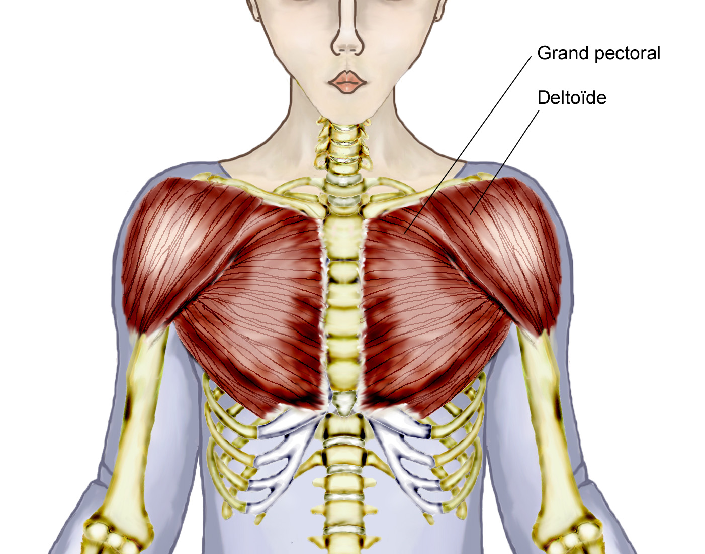
Overview Of Chest Muscles from www.modernheal.com Diagram shoulder muscles human anatomy shoulder muscles amazing neck and shoulder muscles. Printable shoulder muscles diagrams to help you study the muscles structure in human's shoulder.we have five muscle diagrams of the shoulder. Major, subscapularis, coracobrachialis anterior/middle deltoid** spine to the humerus pectoralis. Human anatomy diagrams show internal organs, cells, systems, conditions, symptoms and sickness information and/or tips for healthy living. There are three main muscles in your shoulder: 6 photos of the shoulder muscles labelled diagram. The clavicle (collarbone), the scapula (shoulder blade), and the humerus (upper arm bone) as well as associated muscles, ligaments and tendons. The prerequisite for any treatment in the shoulder region of a patient with pain is a precise and comprehensive picture of the signs and symptoms as they occur during the assessment.
The shoulder muscles produce the characteristic shape of the shoulder and can be classified into two groups:
The shoulder muscles are associated with movements of the upper limb. The primary function of the shoulder girdle is to give strength and range of motion to the arm. Muscular system body anatomy muscle chart anatomy hip muscles anatomy. Deep muscles of shoulder at temple university shoulder anatomy these pictures of this page are about:diagram muscle shoulder joint Tutorials on the shoulder muscles (e.g rotator cuff muscles: It is the major joint connecting the upper limb to the trunk. Shoulder muscles, pictures and descriptions of the movements and attachments. The human shoulder is made up of three bones: Printable shoulder muscles diagrams to help you study the muscles structure in human's shoulder. This diagram depicts shoulder muscle diagram. The anterior deltoid, the lateral deltoid, and the posterior deltoid. Let's start by the anterior view of the diagram. Human anatomy diagrams show internal organs, cells, systems, conditions, symptoms and sickness information and/or tips for healthy living.
The ultimate shoulder workouts anatomy. The anterior deltoid, the lateral deltoid, and the posterior deltoid. Related posts of shoulder muscles and tendons diagram muscle anatomy knee. High quality images of interesting designs, including architectural, graphic, industrial, furniture. The extrinsic muscles of the shoulder include trapezius, latissimus dorsi, levator scapulae, rhomboid major and rhomboid minor.
Coaching Speed: Protracted Shoulder Girdle Part 2 from 3.bp.blogspot.com These muscles aren't as visible as the deltoids, but they are equally (if not more) important. The shoulder joint is formed where the humerus (upper arm bone) fits into the scapula. Printable shoulder muscles diagrams to help you study the muscles structure in human's shoulder. Learn vocabulary, terms and more with flashcards, games and other study tools. The ultimate shoulder workouts anatomy. Muscles allow a person to move muscle tendons in the knee joint and the shoulder joint are crucial in stabilization. The extrinsic muscles of the shoulder include trapezius, latissimus dorsi, levator scapulae, rhomboid major and rhomboid minor. The muscular system consists of various types of muscle that each play a crucial role in the function of the body.
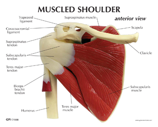
Supraspinatus, infraspinatus, ters minor,.et), using interactive animations and labeled diagrams.
Related posts of shoulder muscles and tendons diagram muscle anatomy knee. It is the major joint connecting the upper limb to the trunk. The shoulder muscles are associated with movements of the upper limb. The shoulder muscles produce the characteristic shape of the shoulder and can be classified into two groups: Human anatomy diagrams show internal organs, cells, systems, conditions, symptoms and sickness information and/or tips for healthy living. The shoulder muscle tissues can be readily injured and therefore being aware of the appropriate strategy is pretty significant when functioning out. Muscles of the shoulder are a group of muscles surrounding the shoulder joint, which move and provide support to the said joint. Muscles of the shoulder and back laminated anatomy chart. The next life study seated female figure, shows the upper part of the pectoralis major positioned flat against the rib cage, with very the muscles of the superficial layer of the back move the shoulder blade (scapula) and upper arm (humerus). This diagram depicts shoulder muscle diagram. Just like the muscle tissues in unique elements of the human physique, even our shoulder muscle tissues are prone to standard put on and tear. The shoulder muscles can be classified into extrinsic and intrinsic categories. 6 photos of the shoulder muscles labelled diagram.
The shoulder joint (glenohumeral joint) is a ball and socket joint between the scapula and the humerus. Although three ligaments protect and surround the shoulder joint, most of its stability comes from the powerful muscles and tendons of the rotator cuff. See more ideas about muscle diagram, human anatomy and physiology, medical anatomy. Let's start by the anterior view of the diagram. Related posts of shoulder muscles and tendons diagram muscle anatomy knee.
Human Shoulder Model with Muscles #1810 for sale | Anatomy Now from www.anatomynow.com The two large main muscles of this. Major, subscapularis, coracobrachialis anterior/middle deltoid** spine to the humerus pectoralis. The shoulder muscle tissues can be readily injured and therefore being aware of the appropriate strategy is pretty significant when functioning out. It is the major joint connecting the upper limb to the trunk. Diagram shoulder muscles human anatomy shoulder muscles amazing neck and shoulder muscles. Muscular system body anatomy muscle chart anatomy hip muscles anatomy. Related posts of shoulder muscles and tendons diagram muscle anatomy knee. Shoulder muscles, shoulder muscles name, shoulder muscles pain, shoulder muscles workout.
Learn vocabulary, terms and more with flashcards, games and other study tools.
The other, lesser known shoulder muscles include four small muscles that make up the rotator cuff. The human shoulder is made up of three bones: Sternum shoulder muscles **muscles on anterior aspect pec. It is the major joint connecting the upper limb to the trunk. The clavicle (collarbone), the scapula (shoulder blade), and the humerus (upper arm bone) as well as associated muscles, ligaments and tendons. The next life study seated female figure, shows the upper part of the pectoralis major positioned flat against the rib cage, with very the muscles of the superficial layer of the back move the shoulder blade (scapula) and upper arm (humerus). The core muscles are those in the abdomen, back, and pelvis, and they. Shoulder muscle and ligament diagram. Muscular system body anatomy muscle chart anatomy hip muscles anatomy. Groin muscles diagram diagram of groin aponeurosis from sscsantry groin project medical. Deep muscles of shoulder at temple university shoulder anatomy these pictures of this page are about:diagram muscle shoulder joint Shoulder muscles, shoulder muscles name, shoulder muscles pain, shoulder muscles workout. The rotator cuff is a complex and delicate structure of.
